Atrioventricular (AV) Canal defect
What is it?
Many terms are used to describe this complex defect. They include atrioventricular (AV) canal, complete AV canal, complete common AV canal, atrioventricular septal defect and endocardial cushion defect.
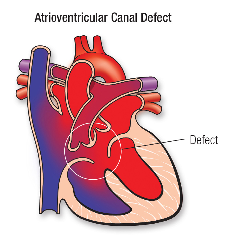
A large hole in the center of the heart affecting all four chambers where they would normally be divided. When a heart is properly divided, the oxygen-rich blood from the lungs does not mix with the oxygen-poor blood from the body. A CAVC allows blood to mix and the chambers and valves to not properly route the blood to each station of circulation.
Atrioventricular (AV) canal defect is a large hole in the center of the heart. It’s located where the wall (septum) between the upper chambers (atria) joins the wall between the lower chambers (ventricles). This septal defect involves both upper and lower chambers. The tricuspid and mitral valves that normally separate the heart’s upper and lower chambers aren’t formed as individual valves. Instead a single large valve forms that crosses the defect in the wall between the two sides of the heart.
What causes it?
In most children, the cause isn’t known. It’s a very common type of heart defect in children with a chromosome problem, Trisomy 21 (Down syndrome). Some children can have other heart defects along with AV canal.
How does it affect the heart?
Normally, the left side of the heart only pumps blood to the body, and the heart’s right side only pumps blood to the lungs. In a child with AV canal defect, blood can travel across the holes from the left heart chambers to the right heart chambers and out into the lung arteries. The extra blood being pumped into the lung arteries makes the heart and lungs work harder and the lungs can become congested.
How does the AV canal defect affect my child?
A child with AV canal defect may breathe faster and harder than normal. Infants may have trouble feeding and growing at a normal rate. Symptoms may not occur until several weeks after birth. High pressure may occur in the blood vessels in the lungs because more blood than normal is being pumped there. Over time this causes permanent damage to the lung blood vessels.
In some infants, the common valve between the upper and lower chambers doesn’t close properly. This lets blood leak backward from the heart’s lower chambers to the upper ones. This leak, called regurgitation or insufficiency, can make the heart work harder, too.
What can be done about the defect?
An AV canal can be fixed. Open-heart surgery is needed to repair the defect. Unlike some other types of septal defects, the AV canal defect can’t close on its own. Medicines may be used temporarily to help with symptoms, but they don’t cure the defect or prevent permanent damage to the lung arteries.
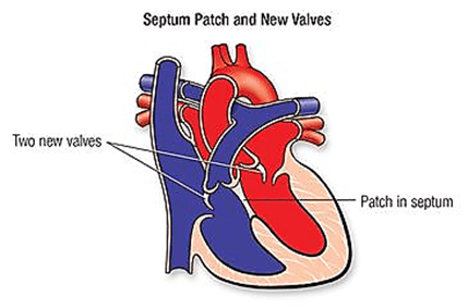
In an infant with severe symptoms or high blood pressure in the lungs, surgery must usually be done in infancy. During the operation, the surgeon closes the large hole with one or two patches. Later the patch will become a permanent part of the heart as the heart’s lining grows over it. The surgeon also divides the single valve between the heart’s upper and lower chambers and makes two separate valves. These will be made as close to normal valves as possible.
If an infant is very ill, or has a defect that may be too complex to repair in infancy, a temporary operation to relieve symptoms and high pressure in the lungs may be needed. This procedure (pulmonary artery banding) narrows the pulmonary artery to reduce the blood flow to the lungs. When the child is older, an operation is done to remove the band and fix the AV canal defect with open-heart surgery.
What activities can my child do?
If the AV canal defect has been closed with surgery, your child may not need any special precautions regarding physical activities and may be able to participate in normal activities without increased risk. Being physically active is healthy for the cardiovascular system, but some children may need to limit their activity. Discuss this with your child’s pediatric cardiologist.
What will my child need in the future?
After surgery your child must be examined regularly by a pediatric cardiologist. More medical or surgical treatment is sometimes needed.
Surgical repair of an AV canal usually restores the blood circulation to normal. However, the reconstructed valve may not work normally. The valve structures can leak or narrow. But, for many children, the long-term outlook is good, and usually no medicines or additional surgery are needed.
What about preventing endocarditis?
Children with AV canal defect may risk endocarditis both before and after repair. Ask about your child’s risk of endocarditis and about your child’s need to take antibiotics before certain dental procedures. See the section on Endocarditis for more information.
If my AV canal defect was closed in childhood, what was done and what can I expect?
The surgery to fix these defects involves patching the ASD and the VSD and repairing the heart valve. To repair the valve, the surgeon divides the single valve between the heart’s upper and lower chambers and makes two separate valves. These are made as close to normal valves as possible. It’s possible that a temporary operation to relieve symptoms and high pressure in the lungs may have been performed before the definitive operation. This procedure (pulmonary artery banding) narrows the pulmonary artery to reduce the blood flow to the lungs. When the child was older, an operation was done to remove the band and fix the AV canal defect with open-heart surgery. Unlike some other types of septal defects, the AV canal defect never closes on its own.
Surgical repair of an AV canal usually restores blood circulation to normal. For many patients, the long-term outlook is good, and no medicines or additional surgery are needed. Because this is a more complicated congenital heart defect, late problems in adults are more common than after an ASD or VSD is closed. As the child grows, the repair may partially break down leading to patch leaks, valve leakage and narrowing of the blood flow channel to the body. These problems may increase the workload of the heart and cause symptoms.
What if the AV canal defect is still present? Should it be repaired in adulthood?
The decision to repair an AV canal defect in adulthood is complicated. It depends on the pressures in the lung and the heart’s pumping function. However, when the pressures aren’t too high and the pump function is good, these defects can be repaired and adult patients are likely to improve. A heart catheterization is almost always required to know whether the defect should be closed. These defects can’t be closed or repaired in the catheterization laboratory, however, because of their location and the need to fix the heart valves.
Problems You May Have
Problems in patients with repaired AV canals depend on whether there are patch leaks and whether there’s a lot of valve regurgitation. Shortness of breath, inability to exercise and swelling in the legs are all signs of heart failure. Abnormal heart rhythms may cause palpitations (skipped or rapid heartbeats) and, rarely, fainting. Some patients may need a pacemaker after the repair if the electrical system has been damaged.
Patients with unrepaired AV canal are often blue (Eisenmenger’s syndrome). Because of valve leaks on the heart’s left side, they’re more likely to have heart failure than other patients with Eisenmenger’s syndrome, due to ASDs and VSDs.
